
You have no items in your shopping cart.
Neuroscience & Cell Signaling
Neurotrophins in Neuronal Development
Understanding their critical roles in cell survival, synaptic plasticity, depression, and neurodegeneration.
1. Functions in Neuronal Development
Neurotrophins are a family of secreted proteins that are absolutely critical for the proper development and long-term maintenance of the nervous system.
Their primary role during development is to actively regulate neuronal populations by promoting the survival of neurons that form appropriate functional connections and inducing programmed cell death (apoptosis) in those that do not. This culling process is essential for establishing correct innervation densities. For instance, in some parts of the vertebrate nervous system, over 50% of all initially generated neurons are deliberately eliminated through this mechanism.
In the adult nervous system, neurotrophins mediate synaptic plasticity, which is fundamental for active learning, memory retention, and overall cognitive function.
Featured Recombinant Proteins
Test direct effects on neuronal cultures to study synaptic plasticity and cell survival pathways with our high-purity Recombinant Human/Mouse BDNF Proteins.
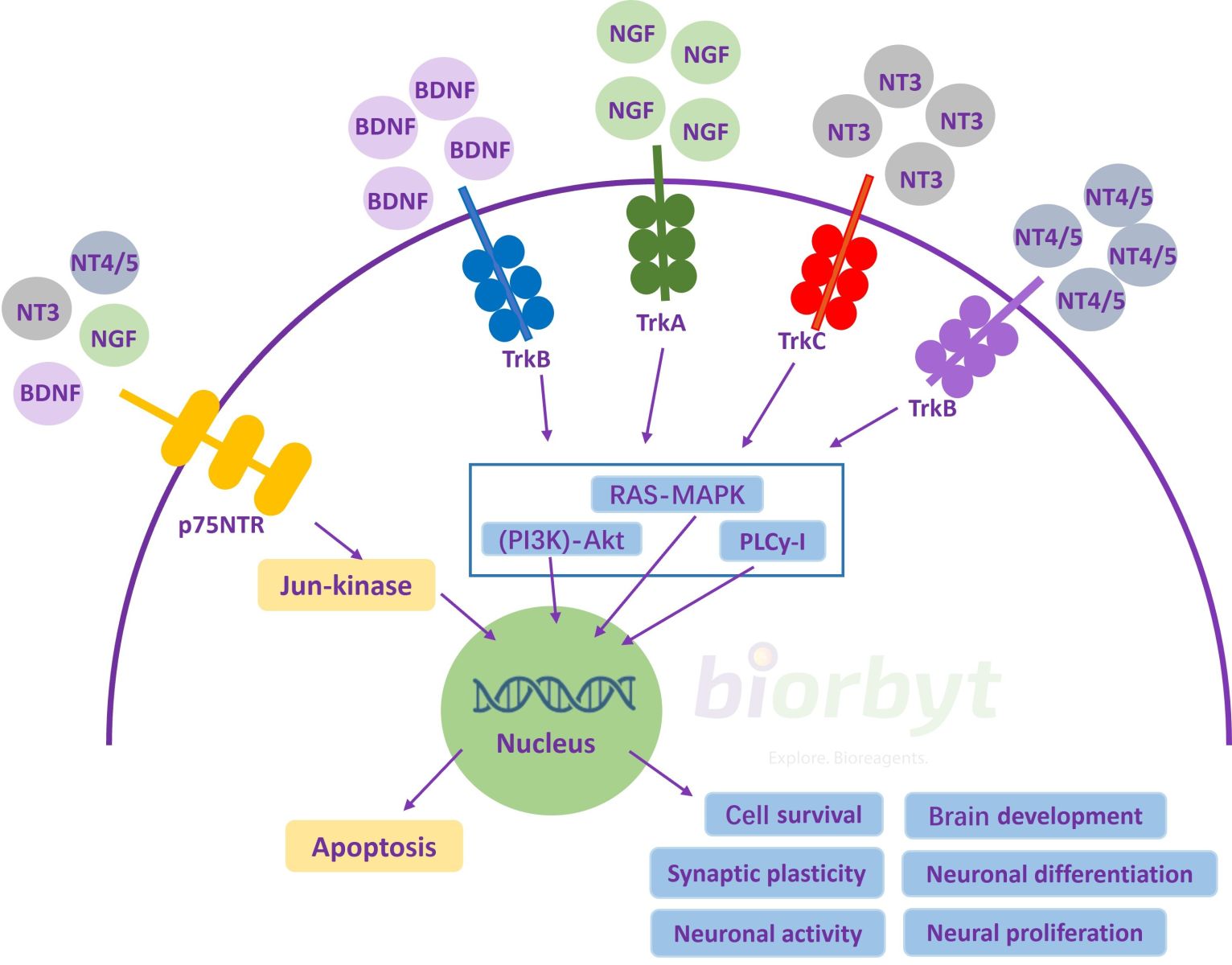
The figure shows the main intracellular signaling pathways and the different effects on the cell.
(Figure from 'Neurotrophin Signaling Impairment by Viral Infections in the Central Nervous System' by Bohmwald, K, et al., Int. Journal of Molecular Sciences, 2022. Licensed under CC BY 4.0.)
2. Core Neurotrophins and Their Receptors
The classical neurotrophin family consists of structurally related proteins that primarily signal through the Tropomyosin receptor kinase (Trk) family of receptor tyrosine kinases.
Nerve Growth Factor (NGF)
The very first neurotrophin to be discovered. NGF is essential for the survival and maintenance of sensory and sympathetic neurons and signals primarily through the TrkA receptor. View NGF Proteins →
Brain-Derived Neurotrophic Factor (BDNF)
BDNF is highly expressed throughout the central nervous system, particularly within the hippocampus, and is a key mediator of synaptic plasticity, learning, and mood regulation. It signals through the TrkB receptor. View BDNF Proteins →
Neurotrophin-3 (NT-3)
NT-3 has a broader range of physiological action. While signaling primarily through the TrkC receptor, it can also activate TrkA and TrkB. It is heavily involved in the development of proprioceptive neurons. View NT-3 Proteins →
Beyond these classical neurotrophins, other prominent growth factors like Fibroblast Growth Factor (FGF) and Ciliary Neurotrophic Factor (CNTF) also exhibit strong neurotrophic activities.
We offer a complete panel of recombinant neurotrophins and highly specific antibodies for TrkA, TrkB, and TrkC, including specialized antibodies that detect critical phosphorylation events (e.g., Anti-phospho-TrkB) to help researchers dissect these complex pathways.
3. Role in Health, Disease, and Therapeutics
Depression and Mood Disorders
Chronic stress is a major clinical risk factor for depression and has been definitively shown to decrease the active expression of BDNF mRNA within the hippocampus. Conversely, a wide range of highly effective antidepressant treatments, from SSRIs to electroconvulsive therapy, robustly increases BDNF expression, strongly suggesting this mechanism is central to their therapeutic action.
- Stress-Induction & Corticosterone Assay Kits: Model chronic stress in vitro and in vivo to study its direct impact on baseline neurotrophin expression.
- BDNF ELISA Kits: Quantify dynamic changes in neurotrophin levels in response to stress or treatment paradigms.
Neurodegenerative Disease
A devastating loss of trophic cellular support is a primary feature of severe neurodegenerative diseases. For example, clinical evidence overwhelmingly points to altered BDNF signaling in the active progression of Alzheimer's disease. This has led to intense modern research into neurotrophin-based therapies. However, the clinical application of neurotrophin proteins has traditionally been severely limited by their poor pharmacokinetic profiles (e.g., a very short half-life) and their physical inability to effectively cross the blood-brain barrier.
- Alzheimer's Disease: A brain disorder that causes progressive memory loss and cognitive decline. It's defined pathologically by the toxic build-up of amyloid-beta protein into plaques between nerve cells and tau protein into tangles inside them, leading to widespread neuron death.
- Parkinson's Disease: A movement disorder causing severe tremors, stiffness, and difficulty with balance. It results directly from the death of brain cells that produce dopamine, and is clinically associated with toxic clumps of alpha-synuclein protein called Lewy bodies.
- Huntington's Disease: An inherited genetic disorder causing uncontrolled movements (chorea) and cognitive decline. It is caused by a mutation in the Huntingtin (Htt) gene, which creates an abnormal protein that forms toxic clumps, leading to the death of neurons.
Nerve Injury and Repair
To accurately assess nerve repair, researchers carefully analyze key protein markers. They reliably visualize overall neuronal structure with Beta III Tubulin, identify active axon regrowth with the primary growth cone marker GAP43, detect inhibitory glial scars using GFAP, and confirm functional myelin recovery by staining for Myelin Basic Protein (MBP). The growth of new axonal or dendritic processes is additionally supported by Neurite Promoting Factors (NPFs) found in the extracellular matrix, such as Laminin and Fibronectin.
| Target Marker | Application & Diagnostic Utility |
|---|---|
| Anti-Beta III Tubulin Antibody | A gold-standard structural marker for identifying neurons and their axons, allowing for the clear, distinct visualization of neuronal morphology and regeneration. |
| Anti-GAP43 Antibody | A highly specific marker for neuronal growth cones—the active, seeking tip of a regenerating axon. Staining for GAP43 highlights exactly where active regrowth is occurring. |
| Anti-GFAP Antibody | A reliable marker for reactive astrocytes. It is heavily used to visualize the formation of the "glial scar," a major physical barrier to axon regeneration in the CNS. |
| Anti-MBP Antibody | Targets Myelin Basic Protein, explicitly allowing for the clear visualization of myelination by Schwann cells (in the PNS) or oligodendrocytes (in the CNS) around regenerated axons. |
4. References
- Oppenheim, R. W. (1991). Cell death during development of the nervous system. Annual Review of Neuroscience, 14(1), 453-501. Link
- Burek, M. J., & Oppenheim, R. W. (1996). Programmed cell death in the developing nervous system. Brain Pathology, 6(4), 427-446. Link
- Poo, M. M. (2001). Neurotrophins as synaptic modulators. Nature Reviews Neuroscience, 2(1), 24-32. Link
- Huang, E. J., & Reichardt, L. F. (2001). Neurotrophins: roles in neuronal development and function. Annual Review of Neuroscience, 24(1), 677-736. Link
- Duman, R. S., & Monteggia, L. M. (2006). A neurotrophic model for stress-related mood disorders. Biological Psychiatry, 59(12), 1116-1127. Link
- Unsicker, K. and Strelau, J. (2000). Functions of transforming growth factor-β isoforms in the nervous system. European Journal of Biochemistry, 267: 6972-6975. Link
- Nibuya, M., Morinobu, S., & Duman, R. S. (1995). Regulation of BDNF and trkB mRNA in rat brain by chronic electroconvulsive seizure and antidepressant drug treatments. Journal of Neuroscience, 15(11), 7539-7547. Link
- Phillips, H. S., et al. (1991). BDNF mRNA is decreased in the hippocampus of individuals with Alzheimer's disease. Neuron, 7(5), 695-702. Link
- Thorne, R. G., & Frey, W. H. (2001). Delivery of neurotrophic factors to the central nervous system: pharmacokinetic considerations. Clinical Pharmacokinetics, 40(12), 907-946. Link
- Mamounas, L. A., et al. (1995). Brain-derived neurotrophic factor promotes the survival and sprouting of serotonergic axons in rat brain. Journal of Neuroscience, 15(12), 7929-7939. Link
- Luckenbill-Edds, L. (1997). Laminin and the mechanism of neuronal outgrowth. Brain Research Reviews, 23(1-2), 1-27. Link