
You have no items in your shopping cart.
.png)
IHC · Spatial Biology · Biomarker Validation
Spatial Biology and IHC: Antibody-Based Biomarker Validation
Spatial biology can reveal new molecular patterns, while IHC, IF and multiplex IF remain essential for protein-level validation, tissue interpretation and translational research.
Spatial biology, single-cell sequencing and AI-driven biology are transforming biomedical research. However, these technologies do not eliminate the need for immunohistochemistry. The future is not replacement. It is integration.
1. Why Spatial Biology Has Become So Important
Spatial biology allows researchers to study molecular signals in their native tissue context. Unlike bulk sequencing, which averages signals across mixed cell populations, spatial methods help reveal where specific transcripts, proteins or cell populations are located within tissue architecture.
This is especially valuable in cancer research, where the position of immune cells, stromal cells and tumor cells can influence disease progression, immune escape and therapeutic response.
| Spatial Biology Question | Research Relevance |
|---|---|
| Where are CD8+ T cells located? | Helps distinguish tumor-infiltrated, excluded or immune-desert phenotypes. |
| Are immune cells close to tumor cells? | Supports analysis of tumor-immune interaction and immune response. |
| Are macrophages enriched in specific regions? | Helps characterize suppressive tumor microenvironment niches. |
| Do biomarker candidates show tissue-specific patterns? | Supports discovery of new diagnostic, prognostic or therapeutic biomarkers. |
2. Why IHC Is Still Essential
Although spatial biology can generate large-scale molecular maps, many important clinical and translational research decisions still depend on established IHC markers. Immunohistochemistry remains widely used because it is cost-effective, fast, interpretable and compatible with routine FFPE tissue workflows.
Research Note: RNA-level spatial discoveries often require antibody-based validation to confirm protein expression, localization and reproducibility.
Cost-effective
IHC is more accessible than high-plex spatial transcriptomics for routine tissue-based studies.
Fast workflow
IHC provides a mature and direct workflow for protein detection in tissue sections.
Protein-level validation
IHC and IF help confirm whether RNA-level discoveries translate into protein expression.
In oncology research, markers such as HER2, ER, PR, Ki-67, PD-L1, CD3 and CD8 continue to rely heavily on high-quality antibodies and tissue-based staining. These markers support research into proliferation, immune infiltration, tumor biology and treatment-related biomarker validation.
3. IHC vs Spatial Biology: Replacement or Integration?
A common misconception is that newer technologies automatically replace established methods. In reality, spatial biology and IHC answer different but complementary questions.
Key Takeaway: Spatial biology is powerful for discovery, but IHC and IF remain essential for protein-level confirmation in real tissue samples.
| Technology | Primary Strength | Role in Research Workflow |
|---|---|---|
| Single-cell sequencing | Identifies cell populations and transcriptional states. | Generates hypotheses and biomarker candidates. |
| Spatial biology | Maps molecular signals within tissue architecture. | Reveals spatial patterns and tissue-level interactions. |
| IHC / IF / multiplex IF | Validates protein expression in tissue context. | Confirms discoveries using antibody-based detection. |
A typical translational workflow may begin with single-cell sequencing, move into spatial biology for tissue mapping, and then return to IHC, IF or multiplex IF for protein-level validation across independent tissue samples.
4. Key IHC and IF Markers for Tumor Microenvironment Research
Tumor microenvironment research often depends on immune, stromal, checkpoint and proliferation markers. These markers are commonly used in IHC, IF, multiplex IF and biomarker validation studies.
| Research Area | Common Markers |
|---|---|
| Immune Checkpoint Research | PD-L1 PD-1 CTLA-4 LAG-3 TIM-3 |
| T Cell Infiltration | CD3 CD4 CD8 FOXP3 |
| Macrophage and Myeloid Biology | CD68 CD163 CD11b |
| Proliferation and Tumor Biology | Ki-67 HER2 EGFR Pan-Cytokeratin |
| Stromal and EMT Research | α-SMA FAP Vimentin E-cadherin N-cadherin |
5. How Biorbyt Supports Spatial Biology Validation
Spatial biology creates new discovery opportunities, but researchers still need reliable reagents to validate those discoveries. Biorbyt supports tissue-based research with antibodies, recombinant proteins, ELISA kits and related tools for IHC, IF, multiplex IF, tumor microenvironment analysis and biomarker validation.
For researchers working with spatial biology data, Biorbyt antibodies can help validate protein expression patterns, confirm biomarker candidates and support follow-up studies using FFPE or frozen tissue sections.
6. Recommended Biorbyt Featured Products
Explore selected Biorbyt antibodies commonly used in IHC, IF, tumor microenvironment research, immune checkpoint studies and biomarker validation.
| Image | Product Details |
|---|---|
|
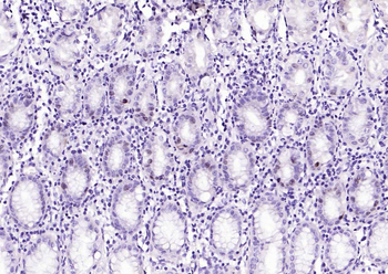
Ki-67 Rabbit Polyclonal Antibody SKU: orb13523 |
|
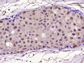
HER2 Mouse Monoclonal Antibody SKU: orb101953 |
|
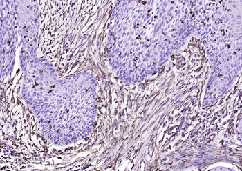
Vimentin Mouse Monoclonal Antibody SKU: orb499662 |
|
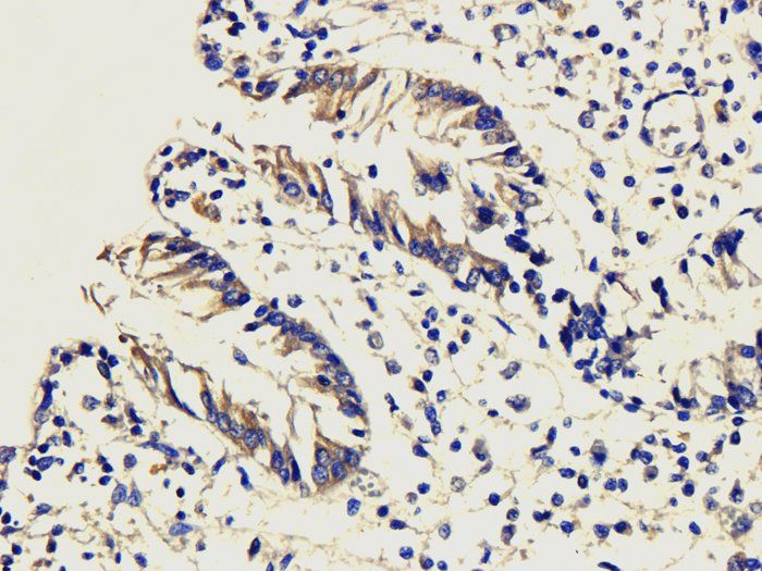
CD4 Rabbit Polyclonal Antibody SKU: orb4830 |
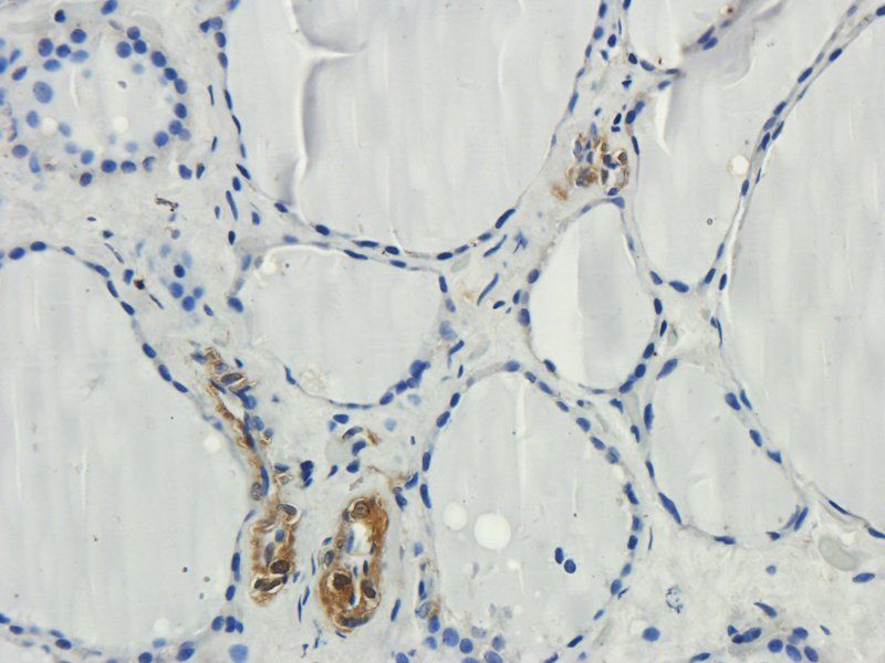
|
CD3 Rabbit Polyclonal Antibody SKU: orb348965 |
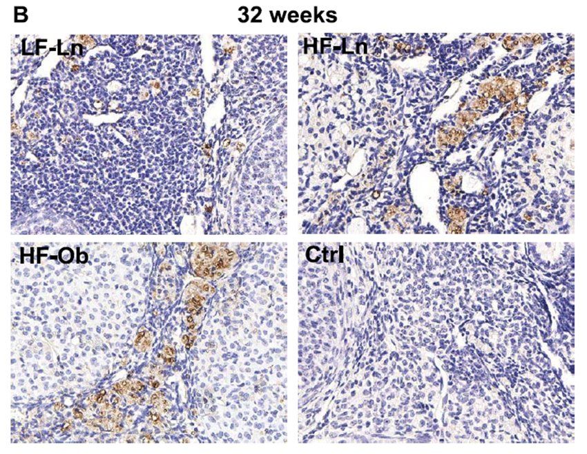
|
CD68 Rabbit Polyclonal Antibody SKU: orb197999 |
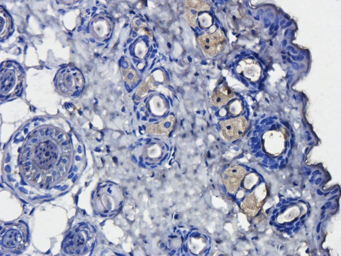
|
CD163 Rabbit Polyclonal Antibody SKU: orb13303 |
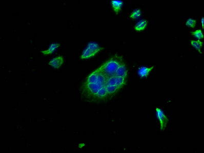
|
PD-L1 Monoclonal Antibody SKU: orb688863 |
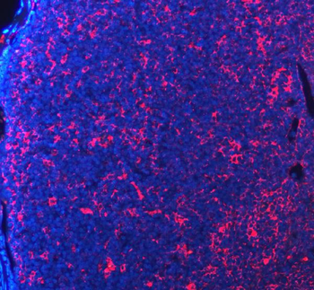
|
PD-1/PDCD1/PD Rabbit Polyclonal Antibody SKU: orb1145869 |
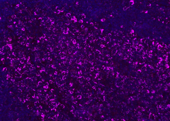
|
CD8B Rabbit Polyclonal Antibody SKU: orb1269 |
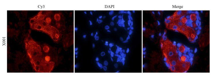
|
FOXP3 Rabbit Polyclonal Antibody SKU: orb34127 |
7. Frequently Asked Questions
Does spatial biology replace IHC?
No. Spatial biology and IHC serve different purposes. Spatial biology is powerful for discovery, while IHC remains essential for protein-level validation, tissue interpretation and biomarker confirmation.
Why is IHC still important in cancer research?
IHC is important because it provides tissue-based protein detection, preserves tissue morphology, supports established biomarker scoring approaches and can be used with FFPE tissue samples.
How is IHC used in biomarker validation?
IHC is commonly used to confirm whether a biomarker is expressed at the protein level, where it is located in tissue and whether its expression pattern is reproducible across independent samples.
What markers are commonly used in tumor microenvironment IHC?
Common tumor microenvironment IHC markers include CD3, CD4, CD8, FOXP3, CD68, CD163, PD-L1, PD-1, Ki-67, HER2, EGFR, α-SMA, FAP and Vimentin.
Why are antibodies important for spatial biology validation?
Spatial biology can identify new cell states, gene expression patterns and biomarker candidates. Antibodies are needed to validate these discoveries at the protein level using IHC, IF or multiplex IF.
8. Quick Search: Popular Markers
Access our validated reagents quickly by browsing through the most commonly searched targets for spatial and immunohistochemistry research.